
Donkey Secondary Antibodies

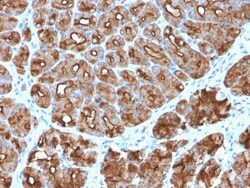
Biotium MUC1 / CA15-3 / EMA / CD227 (Epithelial Marker)(MUC1/2818R), CF647 conjugate, 0.1mg/mL
This MAb reacts with MUC1. The dominant epitope of this MAb has not yet been determined. MUC1 is a large cell surface mucin glycoprotein expressed by most glandular and ductal epithelial cells and some hematopoietic cell lineages. It is expressed on most secretory epithelium, including mammary gland and some hematopoietic cells. It is expressed abundantly in lactating mammary glands and over expressed abundantly in 90% breast carcinomas and metastases. Transgenic MUC1 has been shown to associate with all four c-erbB receptors and localize with c-erbB1 (EGFR) in lactating glands. The MUC1 gene contains seven exons and produces several different alternatively spliced variants. The major expressed form of MUC1 uses all seven exons and is a type 1 transmembrane protein with a large extracellular tandem repeat domain. The tandem repeat domain is highly O glycosylated and alterations in glycosylation have been shown in epithelial cancer cells. Antibody to EMA is useful as a pan-epi

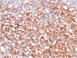
Biotium CD73 (Immuno-Oncology Target)(NT5E/2646), CF647 conjugate, 0.1mg/mL
CD73 is a membrane-bound extracellular enzyme overexpressed in several cancer types. Its expression has been linked to poor prognosis in melanoma, colorectal, gastric, triple negative breast cancer, and to a pro-metastatic phenotype in prostate cancer. Together with CD39, it plays a major role in promoting immunosuppression through the pathway degrading adenosine triphosphate (ATP) into adenosine. Within the tumor microenvironment, ATP promotes immune cell-mediated killing of cancer cells. In contrast, adenosine accumulation causes immune suppression, dysregulation of immune cell infiltrates and stimulates angiogenesis resulting in tumor spreading. CD73 is active on the last step of the degradation pathway, where it is the enzyme that actually degrades AMP into adenosine. CD73-blockade promotes anti-tumor immunity by reducing adenosine accumulation. Accordingly, anti-CD73 mAbs stimulate anti-tumor immunity and reduce tumor metastasis in Mouse tumor models, and could enhance t

Biotium NRF1(NRF1/2609), CF647 conjugate, 0.1mg/mL
The NF-E2 DNA binding protein is composed of two subunits, p45 and MafK, and it regulates expression of globin genes in developing erythroid cells through interaction with Maf recognition elements (MAREs). A family of NF-E2 related proteins, which are collectively known as the Cap 'n' collar (CNC) family and include Nrf1 (also designated TCF11), Nrf2 and Nrf3, are bZIP transcription factors that heterodimerize with Maf proteins to bind MARE sequences. The Nrf proteins also bind the antioxidant response element (ARE) and are implicated in the regulation of detoxification enzymes and the oxidative stress response. They do so by heterodimerizing with Jun family members (c-Jun, JunB and JunD) to activate gene expression, specifically the detoxifying enzyme, NQO1. Nrf2 is widely expressed and is thought to translocate to the nucleus after treatment with xenobiotics and antioxidants, which stimulate its release from a repressor protein Keap1. Nrf3 is highly expressed in placenta, B
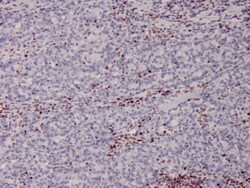
Biotium MSH2 (DNA Mismatch Repair Marker)(MSH2/2622), CF647 conjugate, 0.1mg/mL
Mutations in DNA mismatch repair genes are associated with hereditary nonpolyposis colorectal cancer (HNPCC). Initially, inherited mutations in the MSH2 and MLH1 homologs of the bacterial DNA mismatch repair genes MutS and MutL were found at high frequency in HNPCC and were shown to be associated with microsatellite instability. The demonstration that 10 to 45% of pancreatic, gastric, breast, ovarian and small cell lung cancers also display microsatellite instability has been interpreted to suggest that DNA mismatch repair is not restricted to HNPCC tumors but is a common feature in tumor initiation or progression. Primary antibodies are available purified, or with a selection of fluorescent CF Dyes and other labels. CF Dyes offer exceptional brightness and photostability. Note: Conjugates of blue fluorescent dyes like CF405S and CF405M are not recommended for detecting low abundance targets, because blue dyes have lower fluorescence and can give higher non-specific back
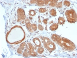
Biotium STAT5B(STAT5B/2611), CF647 conjugate, 0.1mg/mL
Signal transducer and activator of transcription 5A (Stat5a) and Stat5b, which share 96% homology, undergo receptor tyrosine kinase or G protein-coupled receptor-dependent phosphorylation in response to cytokines or growth factors, and then form homo- or heterodimers that translocate to the nucleus, where they initiate transcription. Activation of Stat5a via IL-2, IL-3, IL-7/ GM-CSF, erythropoietin, thrombopoietin and growth hormones influences proliferation, differentiation and apoptosis in lymphohematopoietic cells. Phosphorylation of Stat5a at Ser127/Ser128 and Ser779 are contigent on ErbB-4-mediated activation of Stat5a. Activation of Stat5b via IL-2, IL-4, CSF-1 and growth hormones influences TCR signaling, apoptosis, adult mammary gland development and sexual dimorphism of liver gene expression. Stat5b is the major liver-expressed Stat5 form that has been shown to fuse with the retinoic acid receptor a gene in acute promyelocytic leukemias (APLL). Stat5a/b null mice hav
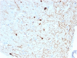
Biotium Calretinin / Calbindin 2 (Mesothelioma Marker)(CALB2/2786), CF647 conjugate, 0.1mg/mL
Calretinin (also known as Calbindin 2) is a member of the troponin C superfamily. Proteins in this family have 6 EF-hand domains which bind to calcium. Calretinin is a vitamin D-dependent calcium-binding protein involved in calcium signaling. It is present in subsets of neurons throughout the brain and spinal chord, including sensory ganglia. Antibodies to calretinin are useful in differentiating mesothelioma from adenocarcinomas of the lung. They can also aid in differentiating adrenal cortical neoplasms from pheochromocytomas. Primary antibodies are available purified, or with a selection of fluorescent CF Dyes and other labels. CF Dyes offer exceptional brightness and photostability. Note: Conjugates of blue fluorescent dyes like CF405S and CF405M are not recommended for detecting low abundance targets, because blue dyes have lower fluorescence and can give higher non-specific background than other dye colors.
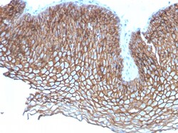
Biotium Desmoglein-3 (Squamous Cell Marker)(DSG3/2837), CF647 conjugate, 0.1mg/mL
Recognizes a protein of 130 kDa, identified as Desmoglein-3 (DSG3). This MAb is highly specific to Desmoglein-3 and does not cross-react with other members of the Desmoglein-family. DSG3 is a calcium-binding transmembrane glycoprotein component of desmosomes in vertebrate epithelial cells. Research has shown that DSG3 has a very high sensitivity (80%) and specificity (100%) in recognizing squamous cell carcinoma (SqCC). Therefore, DSG3 is considered a very important marker for lung SqCC and can be a useful ancillary marker to separate SqCC from other subtypes of lung cancer. Moreover, studies have shown that DSG3 expression in lung SqCC may indicate a poor prognosis. Primary antibodies are available purified, or with a selection of fluorescent CF Dyes and other labels. CF Dyes offer exceptional brightness and photostability. Note: Conjugates of blue fluorescent dyes like CF405S and CF405M are not recommended for detecting low abundance targets, because blue dyes have low

Biotium Napsin A (Lung Adenocarcinoma Marker)(NAPSA/3308), CF647 conjugate, 0.1mg/mL
Napsin is a pepsin-like aspartic proteinase connected with maturation of surfactant protein B. There are two closely related napsins, napsin A and napsin B. Napsin A is expressed as a single chain protein. Immunohistochemical studies revealed high expression levels of napsin A in human lung and kidney but low expression in spleen. Napsin A is expressed in type II pneumocytes and in adenocarcinomas of lung. The high specificity expression of napsin A in adenocarcinomas of lung is useful to distinguish primary lung adenocarcinomas from adenocarcinomas of other organs. Primary antibodies are available purified, or with a selection of fluorescent CF Dyes and other labels. CF Dyes offer exceptional brightness and photostability. Note: Conjugates of blue fluorescent dyes like CF405S and CF405M are not recommended for detecting low abundance targets, because blue dyes have lower fluorescence and can give higher non-specific background than other dye colors.
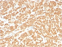
Biotium ICOS-L / ICOS Ligand / B7RP-1 (Immuno-Oncology Target)(ICOSL/3260), CF647 conjugate, 0.1mg/mL
This MAb recognizes a protein of 36 kDa, identified as ICOS-L. It is a ligand for the ICOS receptor that initiates T and B cell proliferation and cytokine secretion. ICOS-L interactions play an essential role in T cell-dependent B cell activation in peripheral lymphoid organs such as spleen and lymph nodes. ICOS-L protein is present in myeloid leukocytes, and by Northern blot there are 2. 4, 3. 0, and 7. 0 kb transcripts in brain, heart, kidney, and liver, with lower expression in colon and thymus, and a 1. 1 kb transcript in leukocytes. Tumor necrosis factor alpha (TNF), granulocyte-macrophage colony-stimulating factor (GM-CSF) and interleukin-4 (IL-4) enhance its expression. LPS-induced up-regulation of ICOS-L is dependent on the MyD88-dependent signaling pathway. Primary antibodies are available purified, or with a selection of fluorescent CF Dyes and other labels. CF Dyes offer exceptional brightness and photostability. Note: Conjugates of blue fluorescent dyes like CF

Biotium Histone H1 (Pan Nuclear Marker)(AE-4), CF647 conjugate, 0.1mg/mL
Please note that this antibody is a recombinant Rabbit version of original anti-histone H1 antibody (Clone AE-4). Because the variable heavy (VH) and variable light (VL) domains are the same, recombinant antibody has the same exact reactivity as the original AE-4 MAb. There are several advantages of producing a recombinant version of a monoclonal antibody. For example, a recombinant antibody is a pure preparation of active immunoglobulin with no contaminating non-functional intact Ig or free light/heavy chains. Secondly, antibody can always be produced, even if hybridoma line is lost. Moreover, it adds the flexibility of converting the antibody to any species, isotype or format. Eukaryotic histones are basic and water-soluble nuclear proteins that form hetero-octameric nucleosome particles by wrapping 146 base pairs of DNA in a left-handed super-helical turn sequentially to form chromosomal fiber. Two molecules of each of the four core histones (H2A, H2B, H3, and H4) form the
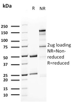
Biotium Uroplakin 1A (Urothelial Differentiation Marker)(UPK1A/2923), CF647 conjugate, 0.1mg/mL
The protein encoded by this gene is a member of the transmembrane 4 superfamily, also known as the tetraspanin family. Most of these members are cell-surface proteins that are characterized by the presence of four hydrophobic domains. The proteins mediate signal transduction events that play a role in the regulation of cell development, activation, growth and motility. This encoded protein is found in the asymmetrical unit membrane (AUM) where it can complex with other transmembrane 4 superfamily proteins. It may play a role in normal bladder epithelial physiology, possibly in regulating membrane permeability of superficial umbrella cells or in stabilizing the apical membrane through AUM/cytoskeletal interactions. The protein may also play a role in tumor suppression. Primary antibodies are available purified, or with a selection of fluorescent CF Dyes and other labels. CF Dyes offer exceptional brightness and photostability. Note: Conjugates of blue fluorescent dyes like
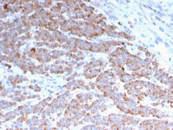
Biotium Cytokeratin 18 (KRT18)(rKRT18/1190), CF568 conjugate, 0.1mg/mL
This MAb reacts with a wide variety of simple epithelia. It does not react with stratified squamous epithelia. It reacts with epithelial tumors of the gastrointestinal tract, lung, breast, pancreas, ovary, and thyroid. Cytokeratin 18, which belongs to the type A (acidic) subfamily of low molecular weight keratins, exists in combination with cytokeratin 8. It was reported that tissues from gastrointestinal tract are positive for both cytokeratin 8 and 18 but do not contain cytokeratin 14. Tissues from gastrointestinal tract, respiratory tract and urogenital tract, as well as endocrine and exocrine tissues and mesothelial cells are positive for cytokeratin 18. Primary antibodies are available purified, or with a selection of fluorescent CF Dyes and other labels. CF Dyes offer exceptional brightness and photostability. Note: Conjugates of blue fluorescent dyes like CF405S and CF405M are not recommended for detecting low abundance targets, because blue dyes have lower fluore
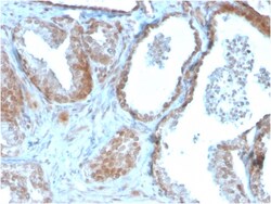
Biotium TIGIT / VSTM3 / VSIG9 (Immune Checkpoint for Cancer)(TIGIT/3106), CF647 conjugate, 0.1mg/mL
TIGIT is a checkpoint inhibitor which binds with high affinity to the poliovirus receptor (PVR), causing increased IL10 secretion, decreased IL12B secretion. TIGIT binding to PVR also causes the suppression of T cell activation by promoting the generation of mature immuno-regulatory dendritic cells. It is expressed at low levels on natural killer (NK) cells, as well as peripheral memory and regulatory CD4 T cells. At the protein level, it is upregulated following the activation of these cells. Functionally, TIGIT is similar to CTLA4. The ligands for TIGIT include CD155 (signal abrogation) and CD226 (signal stimulation). It has been demonstrated to be upregulated on T cells in many cancers and is a immuno-oncology target for therapy. Primary antibodies are available purified, or with a selection of fluorescent CF Dyes and other labels. CF Dyes offer exceptional brightness and photostability. Note: Conjugates of blue fluorescent dyes like CF405S and CF405M are not recomme

Biotium ERCC1 / RAD10 (Tumor Progression Marker)(ERCC1/2683), CF647 conjugate, 0.1mg/mL
Recognizes a protein of 110 kDa, identified as Excision Repair Cross Complementing 1 (ERCC1). It is a mammalian nucleotide excision repair (NER) enzyme involved in repair of damaged DNA. ERCC1 is a homologous to RAD10 in Saccharomyces cerevisiae, which is required in mitotic intrachromosomal recombination and repair. ERCC1 is required in repair of cisplatin-induced DNA adducts and ultraviolet (UV)-induced DNA damage. High expression of ERCC1 has been linked to tumor progression in a variety of cancers including non-small cell lung cancer (NSCLC), squamous cell carcinoma of the head, ovarian cancer and esophageal cancer. Primary antibodies are available purified, or with a selection of fluorescent CF Dyes and other labels. CF Dyes offer exceptional brightness and photostability. Note: Conjugates of blue fluorescent dyes like CF405S and CF405M are not recommended for detecting low abundance targets, because blue dyes have lower fluorescence and can give higher non-specific
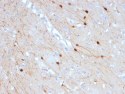
Biotium Calretinin / Calbindin 2 (Mesothelioma Marker)(CALB2/2685), CF647 conjugate, 0.1mg/mL
Calretinin (also known as Calbindin 2) is a member of the troponin C superfamily. Proteins in this family have 6 EF-hand domains which bind to calcium. Calretinin is a vitamin D-dependent calcium-binding protein involved in calcium signaling. It is present in subsets of neurons throughout the brain and spinal chord, including sensory ganglia. Antibodies to calretinin are useful in differentiating mesothelioma from adenocarcinomas of the lung. They can also aid in differentiating adrenal cortical neoplasms from pheochromocytomas. Primary antibodies are available purified, or with a selection of fluorescent CF Dyes and other labels. CF Dyes offer exceptional brightness and photostability. Note: Conjugates of blue fluorescent dyes like CF405S and CF405M are not recommended for detecting low abundance targets, because blue dyes have lower fluorescence and can give higher non-specific background than other dye colors.